Traditionally, we predict cardiac risk from easily-measured factors like age, cholesterol, or blood pressure. A new study in NEJM instead measured “silent” plaque directly through expensive cardiac imaging, and found arterial plaque in 34.6% of men ages 30 to 39. Plaque was also present in 21% of women in their 30s, and 90% of older adults of both sexes.
What’s more interesting is that among 30-to-39-year-olds who had coronary plaque on CT angiography, 41.8% of men and 48.4% of women had a coronary calcium score of zero. The mismatch became less common with age, but it still occurred among people ages 60-70.
How did the study measure this? And what does this all mean? I’ll try to break it aprt below.
Defining plaque
Plaque can be either hard (calcified) or soft (noncalcified). A typical coronary calcium scan measures only hard plaque.
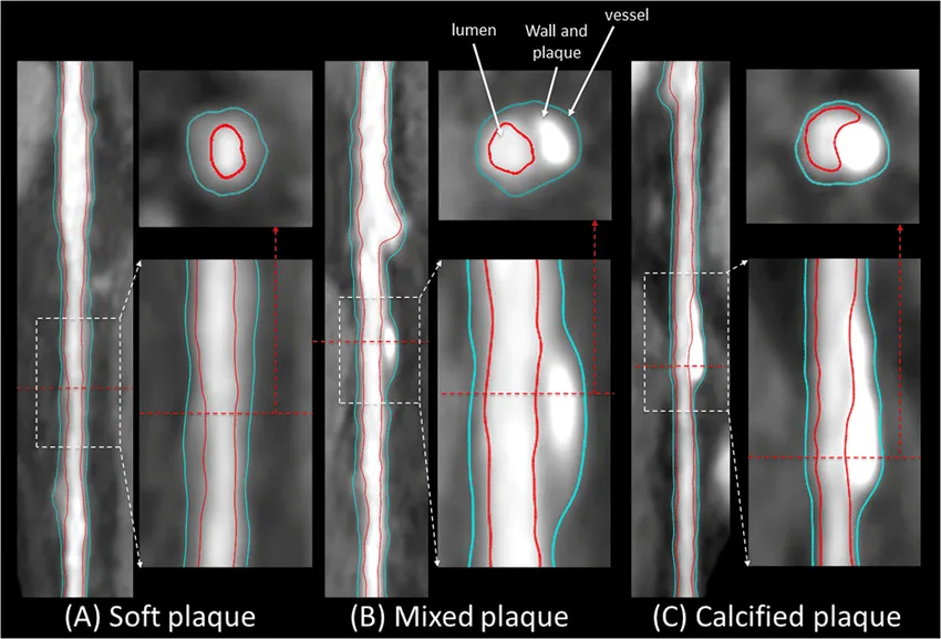 Examples of soft, mixed, and hard (calcified) plaque.
This study used coronary CT angiography (CCTA), which can show noncalcified/soft plaque, for the coronary arteries. For the carotid arteries, researchers u sed three-dimensional vascular ultrasound. Ultrasound identifies a focal thickening or protrusion of the artery wall, and can indirectly estimate plaque volume without radiation or contrast (kind of cool, but a topic for a different article entirely)
Plaque prevalence rises sharply through adult life
Researchers found plaque at every age they studied:
| Age | Men with plaque | Women with plaque |
|---|---|---|
| 18–29 | 8.7% | 6.7% |
| 30–39 | 34.6% | 21.3% |
| 60–70 | 98.1% | 91.9% |
The oldest values are calculated from the reported proportions with no detectable plaque: only 1.9% of men and 8.1% of women ages 60 to 70 had clean imaging in all three arterial territories.
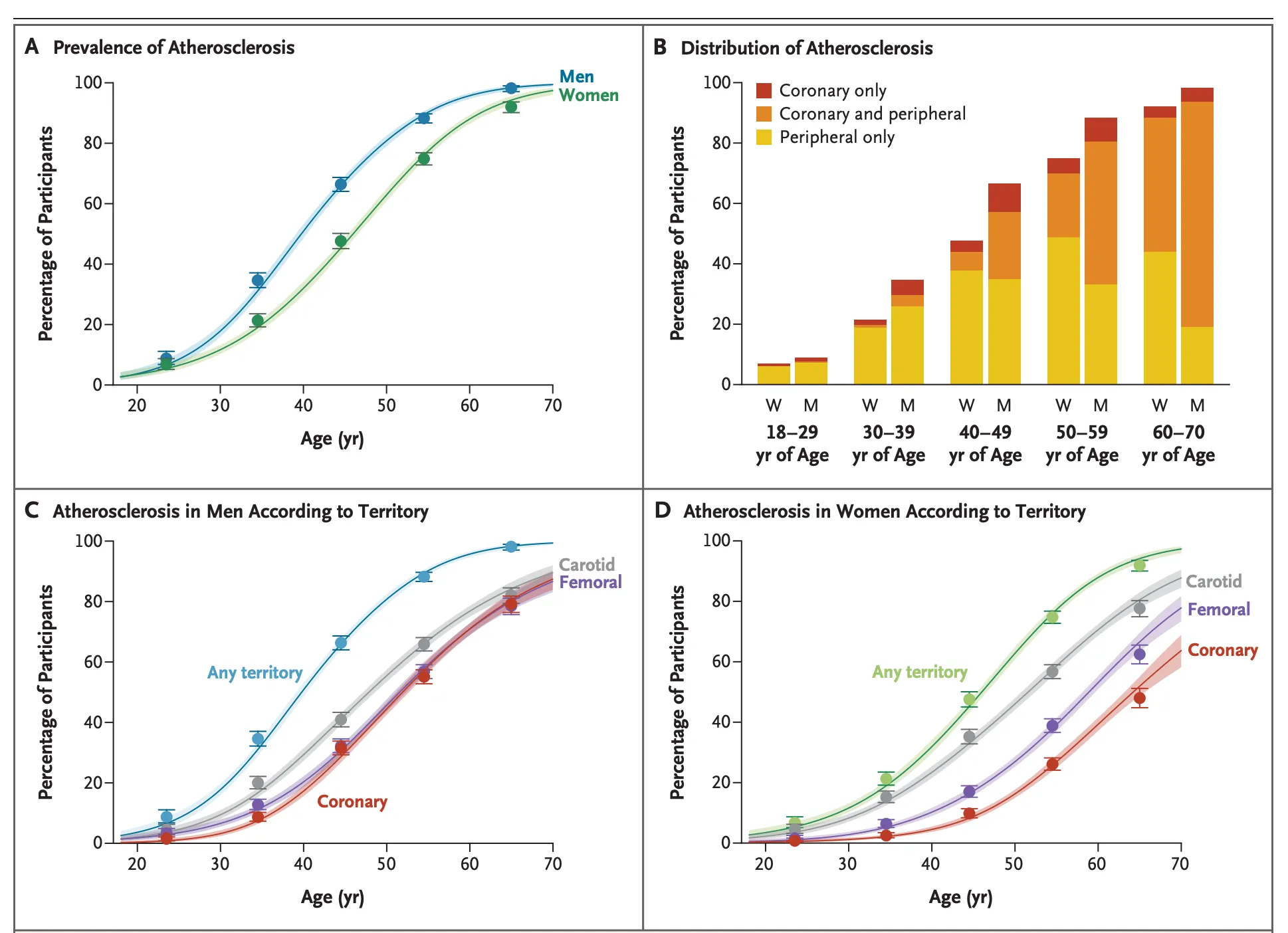 Plaque rises with age. Breakdown of plaque by area. Source: NEJM, 2026.
Where are traditional risk scores most and least accurate?
The researchers calculated a risk score (SCORE2, Europe’s 10-year model for fatal or nonfatal cardiovascular events) at various age ranges, and compared its accuracy at predicting plaque burden:
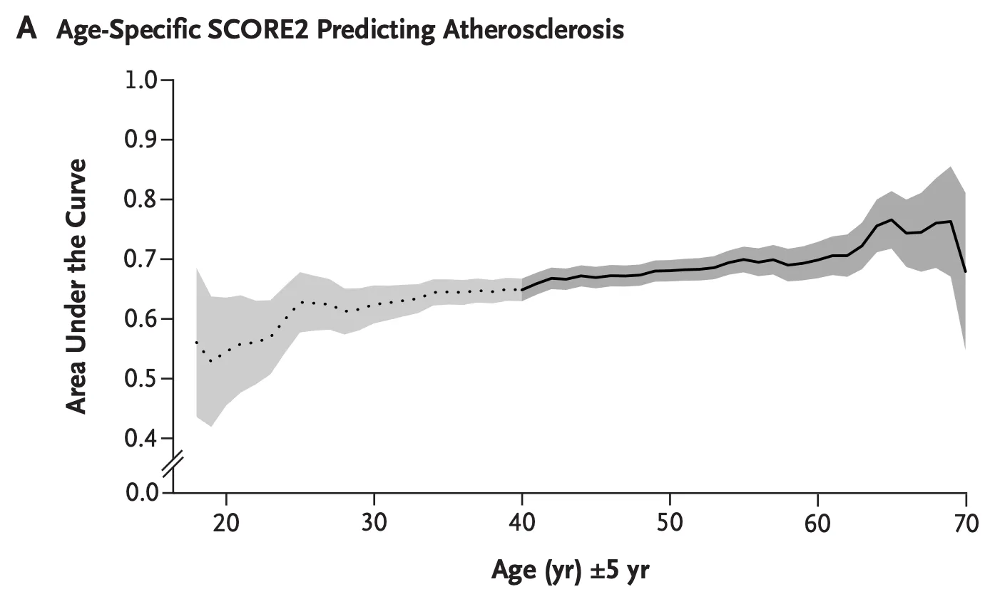 SCORE2’s ability to distinguish participants with and without plaque was modest across most ages. The dotted section is below age 40, where SCORE2 isn’t calibrated. Source: Bundgaard et al., NEJM, 2026.
The risk model was most accurate in older age groups and considerably less acccurate for young age groups. Is this a sign risk models need to be re-calibrated for young people? Or incorporate novel risk factors?
In part, you could argue we’re “abusing” the risk model. Risk models are trained to predict the 10-year risk of a major cardiac event (which is rises with age), which is subtly different the question answered by the scan. But if we belive that heart disease risk accumulates over a lifetime, we do need tools that accurately assess it in the young.
What the study can and can’t tell us
As with many medical studies, you shouldn’t jump up and get a bunch of expensive imaging.
Note that this was a cross-sectional study. Researchers compared different people at different ages rather than watching plaque develop in the same people. Participants came from Denmark and Spain, and healthy-volunteer bias could mean the percentages differ from those in other countries.
The study shows that imaging can find plaque before symptoms, a high calcium score, or a high short-term risk estimate. It doesn’t show that scanning every healthy adult prevents heart attacks. CCTA requires radiation and intravenous contrast, while ultrasound’s value depends on whether its result would change treatment.
For most people, prevention still starts with the factors that drive plaque over time: ApoB or LDL and non-HDL cholesterol, Lp(a), hs-CRP accurate blood-pressure measurement, smoking, diabetes, physical activity, diet, and excess weight. Imaging is most useful when its result could change an uncertain decision, and this study is just one data point toward that direction.
Get your free 30-day heart health guide
Evidence-based steps to optimize your heart health.